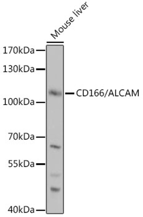
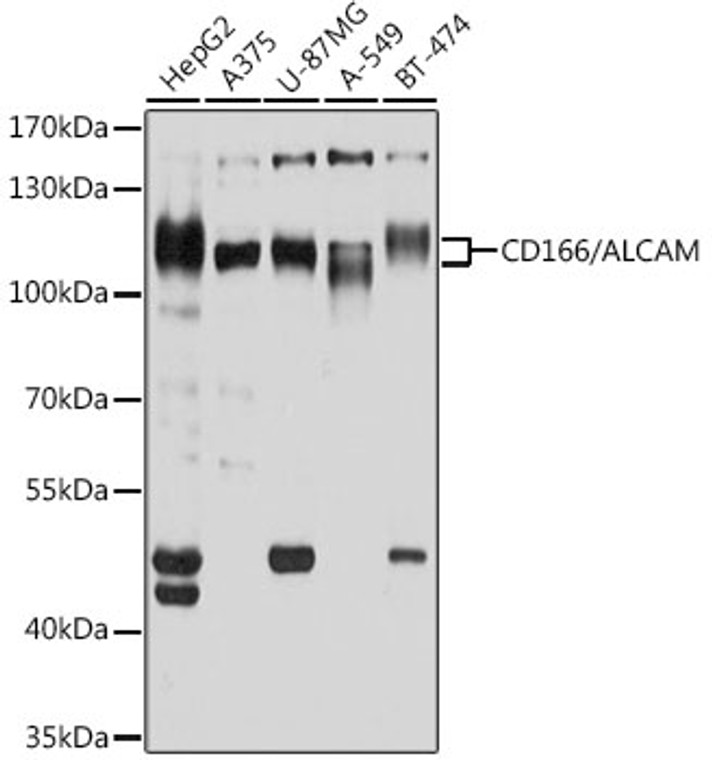
| Host: |
Rabbit |
| Applications: |
WB |
| Reactivity: |
Human/Mouse |
| Note: |
STRICTLY FOR FURTHER SCIENTIFIC RESEARCH USE ONLY (RUO). MUST NOT TO BE USED IN DIAGNOSTIC OR THERAPEUTIC APPLICATIONS. |
| Short Description: |
Rabbit polyclonal antibody anti-ALCAM (28-180) is suitable for use in Western Blot research applications. |
| Clonality: |
Polyclonal |
| Conjugation: |
Unconjugated |
| Isotype: |
IgG |
| Formulation: |
PBS with 0.02% Sodium Azide, 50% Glycerol, pH7.3. |
| Purification: |
Affinity purification |
| Dilution Range: |
WB 1:500-1:2000 |
| Storage Instruction: |
Store at-20°C for up to 1 year from the date of receipt, and avoid repeat freeze-thaw cycles. |
| Gene Symbol: |
ALCAM |
| Gene ID: |
214 |
| Uniprot ID: |
CD166_HUMAN |
| Immunogen Region: |
28-180 |
| Immunogen: |
Recombinant fusion protein containing a sequence corresponding to amino acids 28-180 of human CD166/CD166/ALCAM (NP_001618.2). |
| Immunogen Sequence: |
WYTVNSAYGDTIIIPCRLDV PQNLMFGKWKYEKPDGSPVF IAFRSSTKKSVQYDDVPEYK DRLNLSENYTLSISNARISD EKRFVCMLVTEDNVFEAPTI VKVFKQPSKPEIVSKALFLE TEQLKKLGDCISEDSYPDGN ITWYRNGKVLHPL |
| Tissue Specificity | Detected on hematopoietic stem cells derived from umbilical cord blood. Detected on lymph vessel endothelial cells, skin and tonsil. Detected on peripheral blood monocytes. Detected on monocyte-derived dendritic cells (at protein level). Detected at low levels in spleen, placenta, liver. Expressed by activated T-cells, B-cells, monocytes and thymic epithelial cells. Isoform 1 and isoform 3 are detected in vein and artery endothelial cells, astrocytes, keratinocytes and artery smooth muscle cells. Expressed by neurons in the brain. Restricted expression in tumor cell lines. Detected in highly metastasizing melanoma cell lines. |
| Post Translational Modifications | Glycosylated. |
| Function | Cell adhesion molecule that mediates both heterotypic cell-cell contacts via its interaction with CD6, as well as homotypic cell-cell contacts. Promotes T-cell activation and proliferation via its interactions with CD6. Contributes to the formation and maturation of the immunological synapse via its interactions with CD6. Mediates homotypic interactions with cells that express ALCAM. Acts as a ligand for the LILRB4 receptor, enhancing LILRB4-mediated inhibition of T cell proliferation. Required for normal hematopoietic stem cell engraftment in the bone marrow. Mediates attachment of dendritic cells onto endothelial cells via homotypic interaction. Inhibits endothelial cell migration and promotes endothelial tube formation via homotypic interactions. Required for normal organization of the lymph vessel network. Required for normal hematopoietic stem cell engraftment in the bone marrow. Plays a role in hematopoiesis.required for normal numbers of hematopoietic stem cells in bone marrow. Promotes in vitro osteoblast proliferation and differentiation. Promotes neurite extension, axon growth and axon guidance.axons grow preferentially on surfaces that contain ALCAM. Mediates outgrowth and pathfinding for retinal ganglion cell axons. Isoform 3: Inhibits activities of membrane-bound isoforms by competing for the same interaction partners. Inhibits cell attachment via homotypic interactions. Promotes endothelial cell migration. Inhibits endothelial cell tube formation. |
| Protein Name | Cd166 AntigenActivated Leukocyte Cell Adhesion MoleculeCd Antigen Cd166 |
| Database Links | Reactome: R-HSA-373760 |
| Cellular Localisation | Cell MembraneSingle-Pass Type I Membrane ProteinCell ProjectionAxonDendriteDetected At The Immunological SynapseIEAt The Contact Zone Between Antigen-Presenting Dendritic Cells And T-CellsColocalizes With Cd6 And The Tcr/Cd3 Complex At The Immunological SynapseIsoform 3: Secreted |
| Alternative Antibody Names | Anti-Cd166 Antigen antibodyAnti-Activated Leukocyte Cell Adhesion Molecule antibodyAnti-Cd Antigen Cd166 antibodyAnti-ALCAM antibodyAnti-MEMD antibody |
Information sourced from Uniprot.org
12 months for antibodies. 6 months for ELISA Kits. Please see website T&Cs for further guidance


