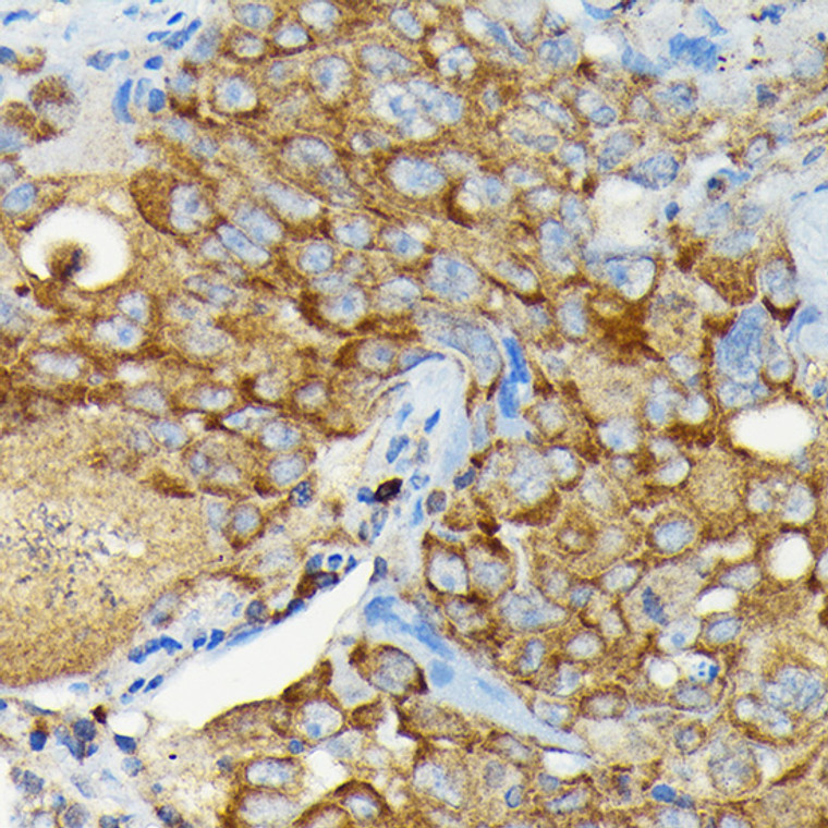
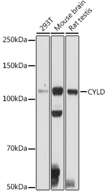
| Host: |
Rabbit |
| Applications: |
WB/IHC |
| Reactivity: |
Human/Mouse/Rat |
| Note: |
STRICTLY FOR FURTHER SCIENTIFIC RESEARCH USE ONLY (RUO). MUST NOT TO BE USED IN DIAGNOSTIC OR THERAPEUTIC APPLICATIONS. |
| Short Description: |
Rabbit polyclonal antibody anti-CYLD (654-953) is suitable for use in Western Blot and Immunohistochemistry research applications. |
| Clonality: |
Polyclonal |
| Conjugation: |
Unconjugated |
| Isotype: |
IgG |
| Formulation: |
PBS with 0.02% Sodium Azide, 50% Glycerol, pH7.3. |
| Purification: |
Affinity purification |
| Dilution Range: |
WB 1:100-1:500IHC-P 1:50-1:200 |
| Storage Instruction: |
Store at-20°C for up to 1 year from the date of receipt, and avoid repeat freeze-thaw cycles. |
| Gene Symbol: |
CYLD |
| Gene ID: |
1540 |
| Uniprot ID: |
CYLD_HUMAN |
| Immunogen Region: |
654-953 |
| Immunogen: |
Recombinant fusion protein containing a sequence corresponding to amino acids 654-953 of human CYLD (NP_001035877.1). |
| Immunogen Sequence: |
TKIMKLRKILEKVEAASGFT SEEKDPEEFLNILFHHILRV EPLLKIRSAGQKVQDCYFYQ IFMEKNEKVGVPTIQQLLEW SFINSNLKFAEAPSCLIIQM PRFGKDFKLFKKIFPSLELN ITDLLEDTPRQCRICGGLAM YECRECYDDPDISAGKIKQF CKTCNTQVHLHPKRLNHKYN PVSLPKDLPDWDWRHGCIPC QNMELFAVLCIETSHYVAFV KYGKDDSAWLFFDSMADRD |
| Tissue Specificity | Detected in fetal brain, testis, and skeletal muscle, and at a lower level in adult brain, leukocytes, liver, heart, kidney, spleen, ovary and lung. Isoform 2 is found in all tissues except kidney. |
| Post Translational Modifications | Ubiquitinated. Polyubiquitinated in hepatocytes treated with palmitic acid. Ubiquitination is mediated by E3 ligase TRIM47 and leads to proteasomal degradation. Phosphorylated on several serine residues by IKKA and/or IKKB in response to immune stimuli. Phosphorylation requires IKBKG. Phosphorylation abolishes TRAF2 deubiquitination, interferes with the activation of Jun kinases, and strongly reduces CD40-dependent gene activation by NF-kappa-B. |
| Function | Deubiquitinase that specifically cleaves 'Lys-63'- and linear 'Met-1'-linked polyubiquitin chains and is involved in NF-kappa-B activation and TNF-alpha-induced necroptosis. Negatively regulates NF-kappa-B activation by deubiquitinating upstream signaling factors. Contributes to the regulation of cell survival, proliferation and differentiation via its effects on NF-kappa-B activation. Negative regulator of Wnt signaling. Inhibits HDAC6 and thereby promotes acetylation of alpha-tubulin and stabilization of microtubules. Plays a role in the regulation of microtubule dynamics, and thereby contributes to the regulation of cell proliferation, cell polarization, cell migration, and angiogenesis. Required for normal cell cycle progress and normal cytokinesis. Inhibits nuclear translocation of NF-kappa-B. Plays a role in the regulation of inflammation and the innate immune response, via its effects on NF-kappa-B activation. Dispensable for the maturation of intrathymic natural killer cells, but required for the continued survival of immature natural killer cells. Negatively regulates TNFRSF11A signaling and osteoclastogenesis. Involved in the regulation of ciliogenesis, allowing ciliary basal bodies to migrate and dock to the plasma membrane.this process does not depend on NF-kappa-B activation. Ability to remove linear ('Met-1'-linked) polyubiquitin chains regulates innate immunity and TNF-alpha-induced necroptosis: recruited to the LUBAC complex via interaction with SPATA2 and restricts linear polyubiquitin formation on target proteins. Regulates innate immunity by restricting linear polyubiquitin formation on RIPK2 in response to NOD2 stimulation. Involved in TNF-alpha-induced necroptosis by removing linear ('Met-1'-linked) polyubiquitin chains from RIPK1, thereby regulating the kinase activity of RIPK1. Negatively regulates intestinal inflammation by removing 'Lys-63' linked polyubiquitin chain of NLRP6, thereby reducing the interaction between NLRP6 and PYCARD/ASC and formation of the NLRP6 inflammasome. Removes 'Lys-63' linked polyubiquitin chain of MAP3K7, which inhibits phosphorylation and blocks downstream activation of the JNK-p38 kinase cascades. Removes also 'Lys-63'-linked polyubiquitin chains of MAP3K1 and MA3P3K3, which inhibit their interaction with MAP2K1 and MAP2K2. |
| Protein Name | Ubiquitin Carboxyl-Terminal Hydrolase CyldDeubiquitinating Enzyme CyldUbiquitin Thioesterase CyldUbiquitin-Specific-Processing Protease Cyld |
| Database Links | Reactome: R-HSA-168638Reactome: R-HSA-5357786Reactome: R-HSA-5357905Reactome: R-HSA-5357956Reactome: R-HSA-5689880Reactome: R-HSA-936440 |
| Cellular Localisation | CytoplasmPerinuclear RegionCytoskeletonCell MembranePeripheral Membrane ProteinCytoplasmic SideMicrotubule Organizing CenterCentrosomeSpindleCilium Basal BodyDetected At The Microtubule Cytoskeleton During InterphaseDetected At The Midbody During TelophaseDuring MetaphaseIt Remains Localized To The Centrosome But Is Also Present Along The Spindle |
| Alternative Antibody Names | Anti-Ubiquitin Carboxyl-Terminal Hydrolase Cyld antibodyAnti-Deubiquitinating Enzyme Cyld antibodyAnti-Ubiquitin Thioesterase Cyld antibodyAnti-Ubiquitin-Specific-Processing Protease Cyld antibodyAnti-CYLD antibodyAnti-CYLD1 antibodyAnti-KIAA0849 antibodyAnti-HSPC057 antibody |
Information sourced from Uniprot.org
12 months for antibodies. 6 months for ELISA Kits. Please see website T&Cs for further guidance


