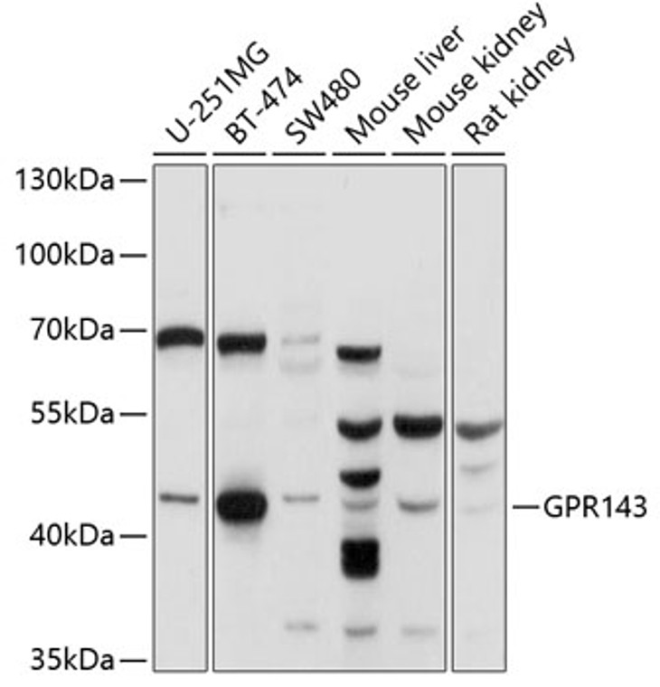
| Host: |
Rabbit |
| Applications: |
WB |
| Reactivity: |
Human/Mouse/Rat |
| Note: |
STRICTLY FOR FURTHER SCIENTIFIC RESEARCH USE ONLY (RUO). MUST NOT TO BE USED IN DIAGNOSTIC OR THERAPEUTIC APPLICATIONS. |
| Short Description: |
Rabbit polyclonal antibody anti-GPR143 (314-404) is suitable for use in Western Blot research applications. |
| Clonality: |
Polyclonal |
| Conjugation: |
Unconjugated |
| Isotype: |
IgG |
| Formulation: |
PBS with 0.02% Sodium Azide, 50% Glycerol, pH7.3. |
| Purification: |
Affinity purification |
| Dilution Range: |
WB 1:1000-1:2000 |
| Storage Instruction: |
Store at-20°C for up to 1 year from the date of receipt, and avoid repeat freeze-thaw cycles. |
| Gene Symbol: |
GPR143 |
| Gene ID: |
4935 |
| Uniprot ID: |
GP143_HUMAN |
| Immunogen Region: |
314-404 |
| Immunogen: |
Recombinant fusion protein containing a sequence corresponding to amino acids 314-404 of human GPR143 (NP_000264.2). |
| Immunogen Sequence: |
TGCSLGFQSPRKEIQWESLT TSAAEGAHPSPLMPHENPAS GKVSQVGGQTSDEALSMLSE GSDASTIEIHTASESCNKNE GDPALPTHGDL |
| Tissue Specificity | Expressed at high levels in the retina, including the retinal pigment epithelium (RPE), and in melanocytes. Weak expression is observed in brain and adrenal gland. |
| Post Translational Modifications | Glycosylated. Phosphorylated. |
| Function | Receptor for tyrosine, L-DOPA and dopamine. After binding to L-DOPA, stimulates Ca(2+) influx into the cytoplasm, increases secretion of the neurotrophic factor SERPINF1 and relocalizes beta arrestin at the plasma membrane.this ligand-dependent signaling occurs through a G(q)-mediated pathway in melanocytic cells. Its activity is mediated by G proteins which activate the phosphoinositide signaling pathway. Also plays a role as an intracellular G protein-coupled receptor involved in melanosome biogenesis, organization and transport. |
| Protein Name | G-Protein Coupled Receptor 143Ocular Albinism Type 1 Protein |
| Database Links | Reactome: R-HSA-375280Reactome: R-HSA-416476 |
| Cellular Localisation | Melanosome MembraneMulti-Pass Membrane ProteinLysosome MembraneApical Cell MembraneDistributed Throughout The Endo-Melanosomal System But Most Of Endogenous Protein Is Localized In Unpigmented Stage Ii MelanosomesIts Expression On The Apical Cell Membrane Is Sensitive To Tyrosine |
| Alternative Antibody Names | Anti-G-Protein Coupled Receptor 143 antibodyAnti-Ocular Albinism Type 1 Protein antibodyAnti-GPR143 antibodyAnti-OA1 antibody |
Information sourced from Uniprot.org
12 months for antibodies. 6 months for ELISA Kits. Please see website T&Cs for further guidance


