| Host: |
Rabbit |
| Applications: |
WB/IHC/IF/ELISA |
| Reactivity: |
Human/Mouse/Rat |
| Note: |
STRICTLY FOR FURTHER SCIENTIFIC RESEARCH USE ONLY (RUO). MUST NOT TO BE USED IN DIAGNOSTIC OR THERAPEUTIC APPLICATIONS. |
| Short Description: |
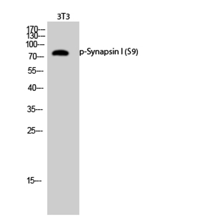
Rabbit polyclonal antibody anti-Phospho-Synapsin-1-Ser9 (3-52 aa) is suitable for use in Western Blot, Immunohistochemistry, Immunofluorescence and ELISA research applications. |
| Clonality: |
Polyclonal |
| Conjugation: |
Unconjugated |
| Isotype: |
IgG |
| Formulation: |
Liquid in PBS containing 50% Glycerol, 0.5% BSA and 0.02% Sodium Azide. |
| Purification: |
The antibody was affinity-purified from rabbit antiserum by affinity-chromatography using epitope-specific immunogen. |
| Concentration: |
1 mg/mL |
| Dilution Range: |
WB 1:500-1:2000IHC 1:100-1:300IF 1:200-1:1000ELISA 1:20000 |
| Storage Instruction: |
Store at-20°C for up to 1 year from the date of receipt, and avoid repeat freeze-thaw cycles. |
| Gene Symbol: |
SYN1 |
| Gene ID: |
6853 |
| Uniprot ID: |
SYN1_HUMAN |
| Immunogen Region: |
3-52 aa |
| Specificity: |
Phospho-Synapsin I (S9) Polyclonal Antibody detects endogenous levels of Synapsin I protein only when phosphorylated at S9. |
| Immunogen: |
The antiserum was produced against synthesized peptide derived from the human Synapsin around the phosphorylation site of Ser9 at the amino acid range 3-52 |
| Function | Neuronal phosphoprotein that coats synaptic vesicles, and binds to the cytoskeleton. Acts as a regulator of synaptic vesicles trafficking, involved in the control of neurotransmitter release at the pre-synaptic terminal. Also involved in the regulation of axon outgrowth and synaptogenesis. The complex formed with NOS1 and CAPON proteins is necessary for specific nitric-oxid functions at a presynaptic level. |
| Protein Name | Synapsin-1Brain Protein 4.1Synapsin I |
| Database Links | Reactome: R-HSA-181429Reactome: R-HSA-212676Reactome: R-HSA-9662360 |
| Cellular Localisation | SynapseGolgi ApparatusPresynapseCytoplasmic VesicleSecretory VesicleSynaptic VesicleDissociates From Synaptic Vesicles And Redistributes Into The Axon During Action Potential FiringIn A Step That Precedes Fusion Of Vesicles With The Plasma MembraneReclusters To Presynapses After The Cessation Of Synaptic Activity |
| Alternative Antibody Names | Anti-Synapsin-1 antibodyAnti-Brain Protein 4.1 antibodyAnti-Synapsin I antibodyAnti-SYN1 antibody |
Information sourced from Uniprot.org
12 months for antibodies. 6 months for ELISA Kits. Please see website T&Cs for further guidance


