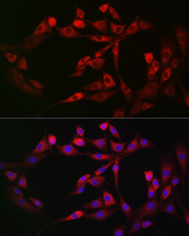
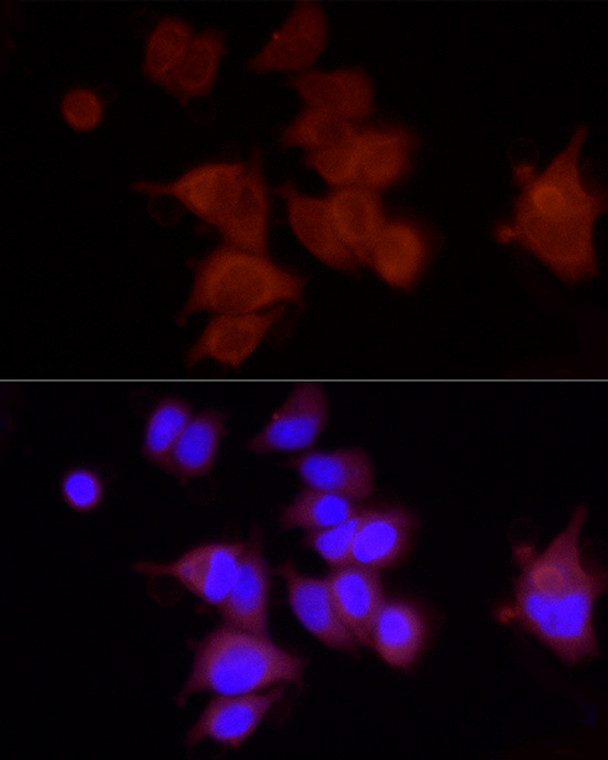
| Host: |
Rabbit |
| Applications: |
WB/IF |
| Reactivity: |
Human/Mouse/Rat |
| Note: |
STRICTLY FOR FURTHER SCIENTIFIC RESEARCH USE ONLY (RUO). MUST NOT TO BE USED IN DIAGNOSTIC OR THERAPEUTIC APPLICATIONS. |
| Short Description: |
Rabbit polyclonal antibody anti-PIEZO1 (2230-2420) is suitable for use in Western Blot and Immunofluorescence research applications. |
| Clonality: |
Polyclonal |
| Conjugation: |
Unconjugated |
| Isotype: |
IgG |
| Formulation: |
PBS with 0.05% Proclin300, 50% Glycerol, pH7.3. |
| Purification: |
Affinity purification |
| Dilution Range: |
WB 1:500-1:1000IF/ICC 1:50-1:200 |
| Storage Instruction: |
Store at-20°C for up to 1 year from the date of receipt, and avoid repeat freeze-thaw cycles. |
| Gene Symbol: |
PIEZO1 |
| Gene ID: |
9780 |
| Uniprot ID: |
PIEZ1_HUMAN |
| Immunogen Region: |
2230-2420 |
| Immunogen: |
Recombinant fusion protein containing a sequence corresponding to amino acids 2230-2420 of human FAM38A/PIEZO1 (NP_001136336.2). |
| Immunogen Sequence: |
PSIIPFTAQAYEELSRQFDP QPLAMQFISQYSPEDIVTAQ IEGSSGALWRISPPSRAQMK RELYNGTADITLRFTWNFQR DLAKGGTVEYANEKHMLALA PNSTARRQLASLLEGTSDQS VVIPNLFPKYIRAPNGPEAN PVKQLQPNEEADYLGVRIQL RREQGAGATGFLEWWVIELQ ECRTDCNLLPM |
| Tissue Specificity | Expressed in numerous tissues. In normal brain, expressed exclusively in neurons, not in astrocytes. In Alzheimer disease brains, expressed in about half of the activated astrocytes located around classical senile plaques. In Parkinson disease substantia nigra, not detected in melanin-containing neurons nor in activated astrocytes. Expressed in erythrocytes (at protein level). Expressed in myoblasts (at protein level). |
| Function | Pore-forming subunit of a mechanosensitive non-specific cation channel. Generates currents characterized by a linear current-voltage relationship that are sensitive to ruthenium red and gadolinium. Plays a key role in epithelial cell adhesion by maintaining integrin activation through R-Ras recruitment to the ER, most probably in its activated state, and subsequent stimulation of calpain signaling. In the kidney, may contribute to the detection of intraluminal pressure changes and to urine flow sensing. Acts as shear-stress sensor that promotes endothelial cell organization and alignment in the direction of blood flow through calpain activation. Plays a key role in blood vessel formation and vascular structure in both development and adult physiology. Acts as sensor of phosphatidylserine (PS) flipping at the plasma membrane and governs morphogenesis of muscle cells. In myoblasts, flippase-mediated PS enrichment at the inner leaflet of plasma membrane triggers channel activation and Ca2+ influx followed by Rho GTPases signal transduction, leading to assembly of cortical actomyosin fibers and myotube formation. |
| Protein Name | Piezo-Type Mechanosensitive Ion Channel Component 1Membrane Protein Induced By Beta-Amyloid TreatmentMibProtein Fam38a |
| Cellular Localisation | Endoplasmic Reticulum MembraneMulti-Pass Membrane ProteinEndoplasmic Reticulum-Golgi Intermediate Compartment MembraneCell MembraneCell ProjectionLamellipodium MembraneIn ErythrocytesLocated In The Plasma MembraneAccumulates At The Leading Apical Lamellipodia Of Endothelial Cells In Response To Shear StressColocalizes With F-Actin And Myh9 At The Actomyosin Cortex In Myoblasts |
| Alternative Antibody Names | Anti-Piezo-Type Mechanosensitive Ion Channel Component 1 antibodyAnti-Membrane Protein Induced By Beta-Amyloid Treatment antibodyAnti-Mib antibodyAnti-Protein Fam38a antibodyAnti-PIEZO1 antibodyAnti-FAM38A antibodyAnti-KIAA0233 antibody |
Information sourced from Uniprot.org
12 months for antibodies. 6 months for ELISA Kits. Please see website T&Cs for further guidance


