| Host: |
Rabbit |
| Applications: |
WB/IHC/IF |
| Reactivity: |
Human/Mouse/Rat |
| Note: |
STRICTLY FOR FURTHER SCIENTIFIC RESEARCH USE ONLY (RUO). MUST NOT TO BE USED IN DIAGNOSTIC OR THERAPEUTIC APPLICATIONS. |
| Short Description: |
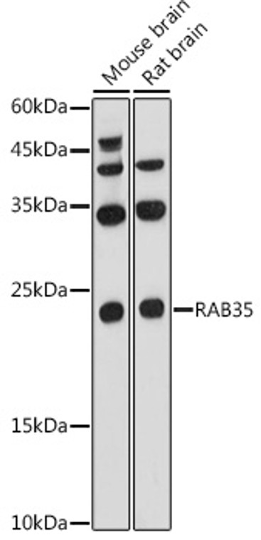
Rabbit polyclonal antibody anti-RAB35 (100-201) is suitable for use in Western Blot, Immunohistochemistry and Immunofluorescence research applications. |
| Clonality: |
Polyclonal |
| Conjugation: |
Unconjugated |
| Isotype: |
IgG |
| Formulation: |
PBS with 0.05% Proclin300, 50% Glycerol, pH7.3. |
| Purification: |
Affinity purification |
| Dilution Range: |
WB 1:500-1:1000IHC-P 1:50-1:200IF/ICC 1:50-1:200 |
| Storage Instruction: |
Store at-20°C for up to 1 year from the date of receipt, and avoid repeat freeze-thaw cycles. |
| Gene Symbol: |
RAB35 |
| Gene ID: |
11021 |
| Uniprot ID: |
RAB35_HUMAN |
| Immunogen Region: |
100-201 |
| Immunogen: |
A synthetic peptide corresponding to a sequence within amino acids 100-201 of human RAB35 (NP_006852.1). |
| Immunogen Sequence: |
KRWLHEINQNCDDVCRILVG NKNDDPERKVVETEDAYKFA GQMGIQLFETSAKENVNVEE MFNCITELVLRAKKDNLAKQ QQQQQNDVVKLTKNSKRKKR CC |
| Post Translational Modifications | AMPylation at Tyr-77 by L.pneumophila DrrA occurs in the switch 2 region and leads to moderate inactivation of the GTPase activity. It appears to prolong the lifetime of the GTP state of RAB1B by restricting access of GTPase effectors to switch 2 and blocking effector-stimulated GTP hydrolysis, thereby rendering RAB35 constitutively active. Phosphocholinated by L.pneumophila AnkX. Both GDP-bound and GTP-bound forms can be phosphocholinated. Phosphocholination inhibits the GEF activity of DENND1A. |
| Function | The small GTPases Rab are key regulators of intracellular membrane trafficking, from the formation of transport vesicles to their fusion with membranes. Rabs cycle between an inactive GDP-bound form and an active GTP-bound form that is able to recruit to membranes different sets of downstream effectors directly responsible for vesicle formation, movement, tethering and fusion. That Rab is involved in the process of endocytosis and is an essential rate-limiting regulator of the fast recycling pathway back to the plasma membrane. During cytokinesis, required for the postfurrowing terminal steps, namely for intercellular bridge stability and abscission, possibly by controlling phosphatidylinositol 4,5-bis phosphate (PIP2) and SEPT2 localization at the intercellular bridge. May indirectly regulate neurite outgrowth. Together with TBC1D13 may be involved in regulation of insulin-induced glucose transporter SLC2A4/GLUT4 translocation to the plasma membrane in adipocytes. |
| Protein Name | Ras-Related Protein Rab-35Gtp-Binding Protein RayRas-Related Protein Rab-1c |
| Database Links | Reactome: R-HSA-8854214Reactome: R-HSA-8873719Reactome: R-HSA-8876198 |
| Cellular Localisation | Cell MembraneLipid-AnchorCytoplasmic SideMembraneClathrin-Coated PitCytoplasmic VesicleClathrin-Coated VesicleEndosomeMelanosomePresent On Sorting Endosomes And Recycling Endosome TubulesTends To Be Enriched In Pip2-Positive Cell Membrane DomainsDuring MitosisAssociated With The Plasma Membrane And Present At The Ingressing Furrow During Early Cytokinesis As Well As At The Intercellular Bridge Later During CytokinesisIdentified In Stage I To Stage Iv Melanosomes |
| Alternative Antibody Names | Anti-Ras-Related Protein Rab-35 antibodyAnti-Gtp-Binding Protein Ray antibodyAnti-Ras-Related Protein Rab-1c antibodyAnti-RAB35 antibodyAnti-RAB1C antibodyAnti-RAY antibody |
Information sourced from Uniprot.org
12 months for antibodies. 6 months for ELISA Kits. Please see website T&Cs for further guidance


