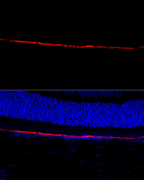
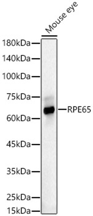
| Host: |
Rabbit |
| Applications: |
WB/IF |
| Reactivity: |
Human/Mouse/Rat |
| Note: |
STRICTLY FOR FURTHER SCIENTIFIC RESEARCH USE ONLY (RUO). MUST NOT TO BE USED IN DIAGNOSTIC OR THERAPEUTIC APPLICATIONS. |
| Short Description: |
Rabbit polyclonal antibody anti-RPE65 (274-533) is suitable for use in Western Blot and Immunofluorescence research applications. |
| Clonality: |
Polyclonal |
| Conjugation: |
Unconjugated |
| Isotype: |
IgG |
| Formulation: |
PBS with 0.02% Sodium Azide, 50% Glycerol, pH7.3. |
| Purification: |
Affinity purification |
| Dilution Range: |
WB 1:1000-1:5000IF/ICC 1:50-1:200 |
| Storage Instruction: |
Store at-20°C for up to 1 year from the date of receipt, and avoid repeat freeze-thaw cycles. |
| Gene Symbol: |
RPE65 |
| Gene ID: |
6121 |
| Uniprot ID: |
RPE65_HUMAN |
| Immunogen Region: |
274-533 |
| Immunogen: |
Recombinant fusion protein containing a sequence corresponding to amino acids 274-533 of human RPE65 (NP_000320.1). |
| Immunogen Sequence: |
NYMDCFESNETMGVWLHIAD KKRKKYLNNKYRTSPFNLFH HINTYEDNGFLIVDLCCWKG FEFVYNYLYLANLRENWEEV KKNARKAPQPEVRRYVLPLN IDKADTGKNLVTLPNTTATA ILCSDETIWLEPEVLFSGPR QAFEFPQINYQKYCGKPYTY AYGLGLNHFVPDRLCKLNVK TKETWVWQEPDSYPSEPIFV SHPDALEEDDGVVLSVVVSP GAGQKPAYLLILNAKDLSE |
| Tissue Specificity | Retina (at protein level). Retinal pigment epithelium specific. |
| Post Translational Modifications | Palmitoylation by LRAT regulates ligand binding specificity.the palmitoylated form (membrane form) specifically binds all-trans-retinyl-palmitate, while the soluble unpalmitoylated form binds all-trans-retinol (vitamin A). |
| Function | Critical isomerohydrolase in the retinoid cycle involved in regeneration of 11-cis-retinal, the chromophore of rod and cone opsins. Catalyzes the cleavage and isomerization of all-trans-retinyl fatty acid esters to 11-cis-retinol which is further oxidized by 11-cis retinol dehydrogenase to 11-cis-retinal for use as visual chromophore. Essential for the production of 11-cis retinal for both rod and cone photoreceptors. Also capable of catalyzing the isomerization of lutein to meso-zeaxanthin an eye-specific carotenoid. The soluble form binds vitamin A (all-trans-retinol), making it available for LRAT processing to all-trans-retinyl ester. The membrane form, palmitoylated by LRAT, binds all-trans-retinyl esters, making them available for IMH (isomerohydrolase) processing to all-cis-retinol. The soluble form is regenerated by transferring its palmitoyl groups onto 11-cis-retinol, a reaction catalyzed by LRAT. |
| Protein Name | Retinoid IsomerohydrolaseAll-Trans-Retinyl-Palmitate HydrolaseLutein IsomeraseMeso-Zeaxanthin IsomeraseRetinal Pigment Epithelium-Specific 65 Kda ProteinRetinol Isomerase |
| Database Links | Reactome: R-HSA-2453902 |
| Cellular Localisation | CytoplasmCell MembraneLipid-AnchorMicrosome MembraneAttached To The Membrane By A Lipid Anchor When Palmitoylated (Membrane Form)Soluble When UnpalmitoylatedUndergoes Light-Dependent Intracellular Transport To Become More Concentrated In The Central Region Of The Retina Pigment Epithelium Cells |
| Alternative Antibody Names | Anti-Retinoid Isomerohydrolase antibodyAnti-All-Trans-Retinyl-Palmitate Hydrolase antibodyAnti-Lutein Isomerase antibodyAnti-Meso-Zeaxanthin Isomerase antibodyAnti-Retinal Pigment Epithelium-Specific 65 Kda Protein antibodyAnti-Retinol Isomerase antibodyAnti-RPE65 antibody |
Information sourced from Uniprot.org
12 months for antibodies. 6 months for ELISA Kits. Please see website T&Cs for further guidance


