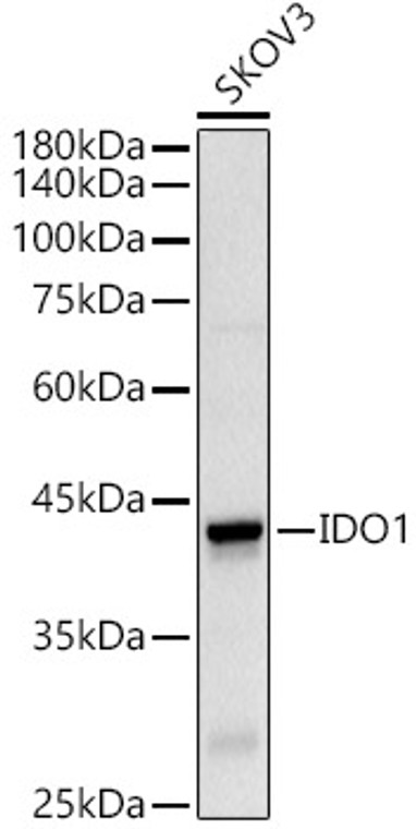
| Host: |
Rabbit |
| Applications: |
WB/IF |
| Reactivity: |
Human/Rat |
| Note: |
STRICTLY FOR FURTHER SCIENTIFIC RESEARCH USE ONLY (RUO). MUST NOT TO BE USED IN DIAGNOSTIC OR THERAPEUTIC APPLICATIONS. |
| Short Description: |
Rabbit polyclonal antibody anti-IDO1 (1-403) is suitable for use in Western Blot and Immunofluorescence research applications. |
| Clonality: |
Polyclonal |
| Conjugation: |
Unconjugated |
| Isotype: |
IgG |
| Formulation: |
PBS with 0.05% Proclin300, 50% Glycerol, pH7.3. |
| Purification: |
Affinity purification |
| Dilution Range: |
WB 1:100-1:500IF/ICC 1:50-1:200 |
| Storage Instruction: |
Store at-20°C for up to 1 year from the date of receipt, and avoid repeat freeze-thaw cycles. |
| Gene Symbol: |
IDO1 |
| Gene ID: |
3620 |
| Uniprot ID: |
I23O1_HUMAN |
| Immunogen Region: |
1-403 |
| Immunogen: |
Recombinant fusion protein containing a sequence corresponding to amino acids 1-403 of human IDO1 (NP_002155.1). |
| Immunogen Sequence: |
MAHAMENSWTISKEYHIDEE VGFALPNPQENLPDFYNDWM FIAKHLPDLIESGQLRERVE KLNMLSIDHLTDHKSQRLAR LVLGCITMAYVWGKGHGDVR KVLPRNIAVPYCQLSKKLEL PPILVYADCVLANWKKKDPN KPLTYENMDVLFSFRDGDCS KGFFLVSLLVEIAAASAIKV IPTVFKAMQMQERDTLLKAL LEIASCLEKALQVFHQIHDH VNPKAFFSVLRIYLSGWKG |
| Tissue Specificity | Expressed in mature dendritic cells located in lymphoid organs (including lymph nodes, spleen, tonsils, Peyers's patches, the gut lamina propria, and the thymic medulla), in some epithelial cells of the female genital tract, as well as in endothelial cells of term placenta and in lung parenchyma. Weakly or not expressed in most normal tissues, but mostly inducible in most tissues. Expressed in more than 50% of tumors, either by tumoral, stromal, or endothelial cells (expression in tumor is associated with a worse clinical outcome). Not overexpressed in tumor-draining lymph nodes. |
| Function | Catalyzes the first and rate limiting step of the catabolism of the essential amino acid tryptophan along the kynurenine pathway. Involved in the peripheral immune tolerance, contributing to maintain homeostasis by preventing autoimmunity or immunopathology that would result from uncontrolled and overreacting immune responses. Tryptophan shortage inhibits T lymphocytes division and accumulation of tryptophan catabolites induces T-cell apoptosis and differentiation of regulatory T-cells. Acts as a suppressor of anti-tumor immunity. Limits the growth of intracellular pathogens by depriving tryptophan. Protects the fetus from maternal immune rejection. |
| Protein Name | Indoleamine 2 -3-Dioxygenase 1Ido-1Indoleamine-Pyrrole 2 -3-Dioxygenase |
| Database Links | Reactome: R-HSA-71240 |
| Cellular Localisation | CytoplasmCytosol |
| Alternative Antibody Names | Anti-Indoleamine 2 -3-Dioxygenase 1 antibodyAnti-Ido-1 antibodyAnti-Indoleamine-Pyrrole 2 -3-Dioxygenase antibodyAnti-IDO1 antibodyAnti-IDO antibodyAnti-INDO antibody |
Information sourced from Uniprot.org
12 months for antibodies. 6 months for ELISA Kits. Please see website T&Cs for further guidance


