• Western blot analysis of extracts of Rat spleen, using IRF1 antibody (STJ110005) at 1:1000 dilution. Secondary antibody: HRP Goat Anti-rabbit IgG (H+L) (STJS000856) at 1:10000 dilution. Lysates/proteins: 25 Mu g per lane. Blocking buffer: 3% non-fat dry milk in TBST. Detection: ECL Basic Kit. Exposure time: 10s.
  • Western blot analysis of extracts of C2C12 cells, using IRF1 antibody (STJ110005) at 1:1000 dilution. C2C12 cells were treated by IFN Gamma (50ng/ml) for 8 hours. Secondary antibody: HRP Goat Anti-rabbit IgG (H+L) (STJS000856) at 1:10000 dilution. Lysates/proteins: 25 Mu g per lane. Blocking buffer: 3% non-fat dry milk in TBST. Detection: ECL Basic Kit. Exposure time: 10s.
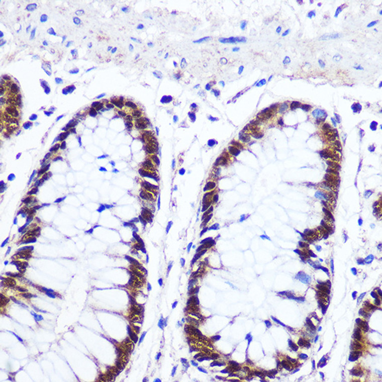
  • Immunohistochemistry analysis of paraffin-embedded Human colon using IRF1 rabbit polyclonal antibody (STJ110005) at dilution of 1:100 (40x lens). Perform microwave antigen retrieval with 10 mM PBS buffer pH 7. 2 before commencing with immunohistochemistry staining protocol.
  • Immunofluorescence analysis of U-2 OS cells using IRF1 antibody (STJ110005) at dilution of 1:100. Blue: DAPI for nuclear staining.

Anti-IRF1 antibody (181-280) (STJ110005)

SKU:
STJ110005

Shipping:
Free Shipping
Current Stock:
Host: Rabbit
Applications: WB/IHC/IF
Reactivity: Human/Mouse/Rat
Note: STRICTLY FOR FURTHER SCIENTIFIC RESEARCH USE ONLY (RUO). MUST NOT TO BE USED IN DIAGNOSTIC OR THERAPEUTIC APPLICATIONS.
Short Description: Rabbit polyclonal antibody anti-IRF1 (181-280) is suitable for use in Western Blot, Immunohistochemistry and Immunofluorescence research applications.
Clonality: Polyclonal
Conjugation: Unconjugated
Isotype: IgG
Formulation: PBS with 0.05% Proclin300, 50% Glycerol, pH7.3.
Purification: Affinity purification
Dilution Range: WB 1:500-1:1000
IHC-P 1:50-1:100
IF/ICC 1:50-1:100
Storage Instruction: Store at-20°C for up to 1 year from the date of receipt, and avoid repeat freeze-thaw cycles.
Gene Symbol: IRF1
Gene ID: 3659
Uniprot ID: IRF1_HUMAN
Immunogen Region: 181-280
Immunogen: A synthetic peptide corresponding to a sequence within amino acids 181-280 of human IRF1 (NP_002189.1).
Immunogen Sequence: PALSPCAVSSTLPDWHIPVE VVPDSTSDLYNFQVSPMPST SEATTDEDEEGKLPEDIMKL LEQSEWQPTNVDGKGYLLNE PGVQPTSVYGDFSCKEEPEI
Post Translational Modifications Phosphorylated by CK2 and this positively regulates its activity. Sumoylation represses the transcriptional activity and displays enhanced resistance to protein degradation. Sumolyated by UBE2I/UBC9 and SUMO1. Inactivates the tumor suppressor activity. Elevated levels in tumor cells. Major site is Lys-275. Sumoylation is enhanced by PIAS3. Desumoylated by SENP1 in tumor cells and appears to compete with ubiquitination on C-terminal sites. Ubiquitinated. Appears to compete with sumoylation on C-terminal sites.
Function Transcriptional regulator which displays a remarkable functional diversity in the regulation of cellular responses. Regulates transcription of IFN and IFN-inducible genes, host response to viral and bacterial infections, regulation of many genes expressed during hematopoiesis, inflammation, immune responses and cell proliferation and differentiation, regulation of the cell cycle and induction of growth arrest and programmed cell death following DNA damage. Stimulates both innate and acquired immune responses through the activation of specific target genes and can act as a transcriptional activator and repressor regulating target genes by binding to an interferon-stimulated response element (ISRE) in their promoters. Competes with the transcriptional repressor ZBED2 for binding to a common consensus sequence in gene promoters. Its target genes for transcriptional activation activity include: genes involved in anti-viral response, such as IFN-alpha/beta, RIGI, TNFSF10/TRAIL, ZBP1, OAS1/2, PIAS1/GBP, EIF2AK2/PKR and RSAD2/viperin.antibacterial response, such as GBP2, GBP5 and NOS2/INOS.anti-proliferative response, such as p53/TP53, LOX and CDKN1A.apoptosis, such as BBC3/PUMA, CASP1, CASP7 and CASP8.immune response, such as IL7, IL12A/B and IL15, PTGS2/COX2 and CYBB.DNA damage responses and DNA repair, such as POLQ/POLH.MHC class I expression, such as TAP1, PSMB9/LMP2, PSME1/PA28A, PSME2/PA28B and B2M and MHC class II expression, such as CIITA.metabolic enzymes, such as ACOD1/IRG1. Represses genes involved in anti-proliferative response, such as BIRC5/survivin, CCNB1, CCNE1, CDK1, CDK2 and CDK4 and in immune response, such as FOXP3, IL4, ANXA2 and TLR4. Stimulates p53/TP53-dependent transcription through enhanced recruitment of EP300 leading to increased acetylation of p53/TP53. Plays an important role in immune response directly affecting NK maturation and activity, macrophage production of IL12, Th1 development and maturation of CD8+ T-cells. Also implicated in the differentiation and maturation of dendritic cells and in the suppression of regulatory T (Treg) cells development. Acts as a tumor suppressor and plays a role not only in antagonism of tumor cell growth but also in stimulating an immune response against tumor cells.
Protein Name Interferon Regulatory Factor 1
Irf-1
Database Links Reactome: R-HSA-5620971
Reactome: R-HSA-877300
Reactome: R-HSA-909733
Reactome: R-HSA-983231
Cellular Localisation Nucleus
Cytoplasm
Myd88-Associated Irf1 Migrates Into The Nucleus More Efficiently Than Non-Myd88-Associated Irf1
Alternative Antibody Names Anti-Interferon Regulatory Factor 1 antibody
Anti-Irf-1 antibody
Anti-IRF1 antibody

Information sourced from Uniprot.org

12 months for antibodies. 6 months for ELISA Kits. Please see website T&Cs for further guidance