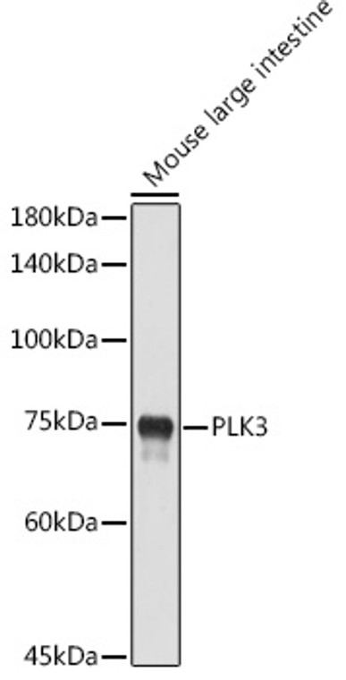
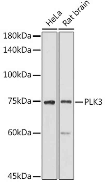
| Host: |
Rabbit |
| Applications: |
WB |
| Reactivity: |
Human/Mouse/Rat |
| Note: |
STRICTLY FOR FURTHER SCIENTIFIC RESEARCH USE ONLY (RUO). MUST NOT TO BE USED IN DIAGNOSTIC OR THERAPEUTIC APPLICATIONS. |
| Short Description: |
Rabbit polyclonal antibody anti-PLK3 (487-646) is suitable for use in Western Blot research applications. |
| Clonality: |
Polyclonal |
| Conjugation: |
Unconjugated |
| Isotype: |
IgG |
| Formulation: |
PBS with 0.02% Sodium Azide, 50% Glycerol, pH7.3. |
| Purification: |
Affinity purification |
| Dilution Range: |
WB 1:500-1:1000 |
| Storage Instruction: |
Store at-20°C for up to 1 year from the date of receipt, and avoid repeat freeze-thaw cycles. |
| Gene Symbol: |
PLK3 |
| Gene ID: |
1263 |
| Uniprot ID: |
PLK3_HUMAN |
| Immunogen Region: |
487-646 |
| Immunogen: |
Recombinant fusion protein containing a sequence corresponding to amino acids 487-646 of human PLK3 (NP_004064.2). |
| Immunogen Sequence: |
VLFNDGTHMALSANRKTVHY NPTSTKHFSFSVGAVPRALQ PQLGILRYFASYMEQHLMKG GDLPSVEEVEVPAPPLLLQW VKTDQALLMLFSDGTVQVNF YGDHTKLILSGWEPLLVTFV ARNRSACTYLASHLRQLGCS PDLRQRLRYALRLLRDRSPA |
| Tissue Specificity | Transcripts are highly detected in placenta, lung, followed by skeletal muscle, heart, pancreas, ovaries and kidney and weakly detected in liver and brain. May have a short half-live. In cells of hematopoietic origin, strongly and exclusively detected in terminally differentiated macrophages. Transcript expression appears to be down-regulated in primary lung tumor. |
| Post Translational Modifications | Phosphorylated in an ATM-dependent manner following DNA damage. Phosphorylated as cells enter mitosis and dephosphorylated as cells exit mitosis. |
| Function | Serine/threonine-protein kinase involved in cell cycle regulation, response to stress and Golgi disassembly. Polo-like kinases act by binding and phosphorylating proteins are that already phosphorylated on a specific motif recognized by the POLO box domains. Phosphorylates ATF2, BCL2L1, CDC25A, CDC25C, CHEK2, HIF1A, JUN, p53/TP53, p73/TP73, PTEN, TOP2A and VRK1. Involved in cell cycle regulation: required for entry into S phase and cytokinesis. Phosphorylates BCL2L1, leading to regulate the G2 checkpoint and progression to cytokinesis during mitosis. Plays a key role in response to stress: rapidly activated upon stress stimulation, such as ionizing radiation, reactive oxygen species (ROS), hyperosmotic stress, UV irradiation and hypoxia. Involved in DNA damage response and G1/S transition checkpoint by phosphorylating CDC25A, p53/TP53 and p73/TP73. Phosphorylates p53/TP53 in response to reactive oxygen species (ROS), thereby promoting p53/TP53-mediated apoptosis. Phosphorylates CHEK2 in response to DNA damage, promoting the G2/M transition checkpoint. Phosphorylates the transcription factor p73/TP73 in response to DNA damage, leading to inhibit p73/TP73-mediated transcriptional activation and pro-apoptotic functions. Phosphorylates HIF1A and JUN is response to hypoxia. Phosphorylates ATF2 following hyperosmotic stress in corneal epithelium. Also involved in Golgi disassembly during the cell cycle: part of a MEK1/MAP2K1-dependent pathway that induces Golgi fragmentation during mitosis by mediating phosphorylation of VRK1. May participate in endomitotic cell cycle, a form of mitosis in which both karyokinesis and cytokinesis are interrupted and is a hallmark of megakaryocyte differentiation, via its interaction with CIB1. |
| Protein Name | Serine/Threonine-Protein Kinase Plk3Cytokine-Inducible Serine/Threonine-Protein KinaseFgf-Inducible KinasePolo-Like Kinase 3Plk-3Proliferation-Related Kinase |
| Database Links | Reactome: R-HSA-6804115Reactome: R-HSA-6804756 |
| Cellular Localisation | CytoplasmNucleusNucleolusGolgi ApparatusCytoskeletonMicrotubule Organizing CenterCentrosomeTranslocates To The Nucleus Upon Cisplatin TreatmentLocalizes To The Golgi Apparatus During InterphaseAccording To A ReportPlk3 Localizes Only In The Nucleolus And Not In The CentrosomeOr In Any Other Location In The CytoplasmThe Discrepancies In Results May Be Explained By The Plk3 Antibody SpecificityBy Cell Line-Specific Expression Or Post-Translational Modifications |
| Alternative Antibody Names | Anti-Serine/Threonine-Protein Kinase Plk3 antibodyAnti-Cytokine-Inducible Serine/Threonine-Protein Kinase antibodyAnti-Fgf-Inducible Kinase antibodyAnti-Polo-Like Kinase 3 antibodyAnti-Plk-3 antibodyAnti-Proliferation-Related Kinase antibodyAnti-PLK3 antibodyAnti-CNK antibodyAnti-FNK antibodyAnti-PRK antibody |
Information sourced from Uniprot.org
12 months for antibodies. 6 months for ELISA Kits. Please see website T&Cs for further guidance


![Western blot analysis of lysates from wild type (WT) and MST1/STK4 knockout (KO) HeLa cells, using [KO Validated] MST1/STK4 Rabbit polyclonal antibody (STJ11100073) at 1:1000 dilution. Secondary antibody: HRP Goat Anti-Rabbit IgG (H+L) (STJS000856) at 1:10000 dilution. Lysates/proteins: 25 Mu g per lane. Blocking buffer: 3% nonfat dry milk in TBST. Detection: ECL Basic Kit. Exposure time: 1s. Western blot analysis of lysates from wild type (WT) and MST1/STK4 knockout (KO) HeLa cells, using [KO Validated] MST1/STK4 Rabbit polyclonal antibody (STJ11100073) at 1:1000 dilution. Secondary antibody: HRP Goat Anti-Rabbit IgG (H+L) (STJS000856) at 1:10000 dilution. Lysates/proteins: 25 Mu g per lane. Blocking buffer: 3% nonfat dry milk in TBST. Detection: ECL Basic Kit. Exposure time: 1s.](https://cdn11.bigcommerce.com/s-zso2xnchw9/images/stencil/300x300/products/89143/357593/STJ11100073_1__61039.1713121731.jpg?c=1)