

. 2, STJ95402")

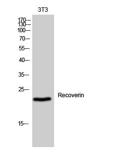
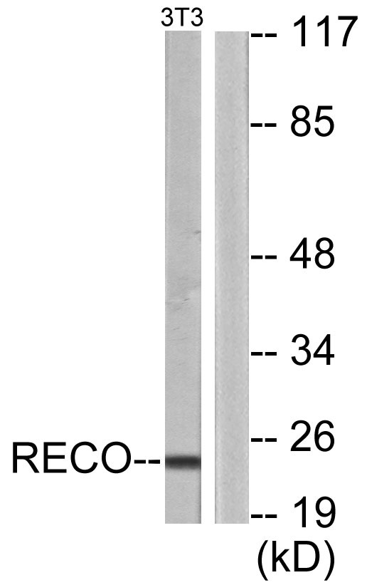
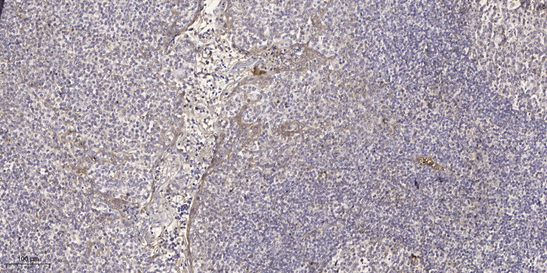
Anti-RCVRN antibody (107-156 aa) (STJ95402)
SPECIFICATIONS
ClonalityPolyclonal
HostRabbit
ConjugationUnconjugated
IsotypeIgG
ImmunogenThe antiserum was produced against synthesized peptide derived from the human Recoverin at the amino acid range 107-156
General Information
| Short Description | Rabbit polyclonal anti-Recoverin (107-156 aa) for use in WB, IHC, IF and ELISA in Human and Mouse samples. Datasheet included with dilution recommendations, and related reagents. |
| Applications | WB/IHC/IF/ELISA |
| Host | Rabbit |
| Reactivity | Human/Mouse |
| Note | STRICTLY FOR FURTHER SCIENTIFIC RESEARCH USE ONLY (RUO). MUST NOT TO BE USED IN DIAGNOSTIC OR THERAPEUTIC APPLICATIONS. |
Product Properties
| Clonality | Polyclonal |
| Isotype | IgG |
| Conjugation | Unconjugated |
| Concentration | 1 mg/mL |
| Purification | The antibody was affinity-purified from rabbit antiserum by affinity-chromatography using epitope-specific immunogen. |
| Dilution Range | WB 1:500-1:2000IHC 1:100-1:300IF 1:200-1:1000ELISA 1:20000 |
| Formulation | Liquid in PBS containing 50% Glycerol, 0.5% BSA and 0.02% Sodium Azide. |
| Storage Instruction | Store at-20°C for up to 1 year from the date of receipt, and avoid repeat freeze-thaw cycles. |
Target Information
| Gene Symbol | RCVRN |
| Gene ID | 5957 |
| Uniprot ID | RECO_HUMAN |
| Immunogen | The antiserum was produced against synthesized peptide derived from the human Recoverin at the amino acid range 107-156 |
| Immunogen Region | 107-156 aa |
| Specificity | Recoverin Polyclonal Antibody detects endogenous levels of Recoverin protein. |
Additional Info
| Post Translational Modifications | The N-terminal glycine is linked to one of four different types of acyl groups. The most abundant is myristoleate (14:1), but 14:0, 14:2, and 12:0 acyl residues are also present. The Ca(2+) induced exposure of the myristoyl group, known as the calcium-myristoyl switch, promotes RCVRN binding to the photoreceptor cell membranes only when intracellular Ca(2+) concentration is high. Oxidation on Cys-39 occurs in response to prolonged intense illumination and results in the formation of disulfide homodimers, and to a lesser extent disulfide-linked heterodimers. |
| Function | Acts as a calcium sensor and regulates phototransduction of cone and rod photoreceptor cells. Modulates light sensitivity of cone photoreceptor in dark and dim conditions. In response to high Ca(2+) levels induced by low light levels, prolongs RHO/rhodopsin activation in rod photoreceptor cells by binding to and inhibiting GRK1-mediated phosphorylation of RHO/rhodopsin. Plays a role in scotopic vision/enhances vision in dim light by enhancing signal transfer between rod photoreceptors and rod bipolar cells. Improves rod photoreceptor sensitivity in dim light and mediates response of rod photoreceptors to facilitate detection of change and motion in bright light. |
| Protein Name | RecoverinCancer-Associated Retinopathy ProteinProtein Car |
| Database Links | Reactome: R-HSA-2514859 |
| Cellular Localisation | Photoreceptor Inner SegmentCell ProjectionCiliumPhotoreceptor Outer SegmentPhotoreceptor Outer Segment MembraneLipid-AnchorCytoplasmic SidePerikaryonPrimarily Expressed In The Inner Segments Of Light-Adapted Rod PhotoreceptorsApproximately 10% Of Which Translocates From Photoreceptor Outer Segments Upon Light StimulationTargeting Of Myristoylated Protein To Rod Photoreceptor Outer Segments Is Calcium Dependent |
| Alternative Antibody Names | Anti-Recoverin antibodyAnti-Cancer-Associated Retinopathy Protein antibodyAnti-Protein Car antibodyAnti-RCVRN antibodyAnti-RCV1 antibody |
Information sourced from Uniprot.org